
category
- ALL
- Bilingual Doctor
- Blepharoplasty
- Breast augmentation
- Deep Plane ミニリフト
- Extended Deep Plane Facelift
- HIFU
- Medical Hair Removal
- Rejuran
- Rhinoplasty
- Uncategorized
- お知らせ
- カウンセリング
- くま治療
- しみ
- スキンボトックス
- たるみ
- ヒアルロン酸
- フェイスリフト
- ボトックス
- ボルニューマ
- メンズ美容医療
- わきボトックス
- わき汗
- 乳輪の色素改善
- 他院修正
- 医療アートメイク
- 医療脱毛
- 学会活動
- 新メニュー
- 毛穴
- 沖縄
- 無断キャンセル
- 症例画像
- 眼瞼下垂
- 硬毛化
- 立体脂肪注入
- 美容
- 美容外科学会(JSAPS)
- 肌メンテ
- 肌育
- 肌質改善
- 肝斑
- 脂肪注入
- 自分らしさを消さない
- 裏ハムラ
- 論文
- 豊胸
- 針脱毛
- 骨切り
Under-Eye Bags and Dark Circles: Understanding the Contour Beneath the Eyes
監修医師

MSA美容外科 院長モレロ オースティン誠
1989年、アメリカ生まれ。10歳より沖縄で育つ。日本とアメリカ、双方の文化や価値観に触れてきた経験を生かし、患者様一人ひとりが持つ異なる美意識や希望を尊重した診療を大切にしています。日本語・英語の両方で診療を行い、国内外の知見を取り入れながら、自然で長期的な美しさを目指した美容医療を提供しています。大手美容外科にて約8年間、美容外科・美容皮膚科診療に携わり、2024年にMSA美容外科を開院。
略歴
2018〜2024年 大手美容外科沖縄院 院長
2024年 MSA美容外科 開院
資格・所属学会
日本美容外科学会(JSAS)認定専門医
日本美容外科学会(JSAS)正会員
日本美容外科学会(JSAPS)関連会員
The Rhinoplasty Society 海外会員
American Academy of Cosmetic Surgery(AACS)正会員
日本美容皮膚科学会 正会員
日本美容外科医師会 正会員
Understanding Under-Eye Bags and Dark Circles: The Anatomy Behind a Tired Appearance
“I look tired, even when I feel well.”
This is a common concern among patients seeking treatment for under-eye bags and dark circles. Understanding the anatomy beneath the skin helps explain why these changes develop—and why treatment must be tailored to each person.
Dark circles may reflect pigmentation, thin skin, visible blood vessels, or shadows created by an uneven contour. Surgery primarily addresses the structural component: the relationship between a prominent lower eyelid and the hollow beneath it.
The tear trough ligament: why a groove forms beneath the eye
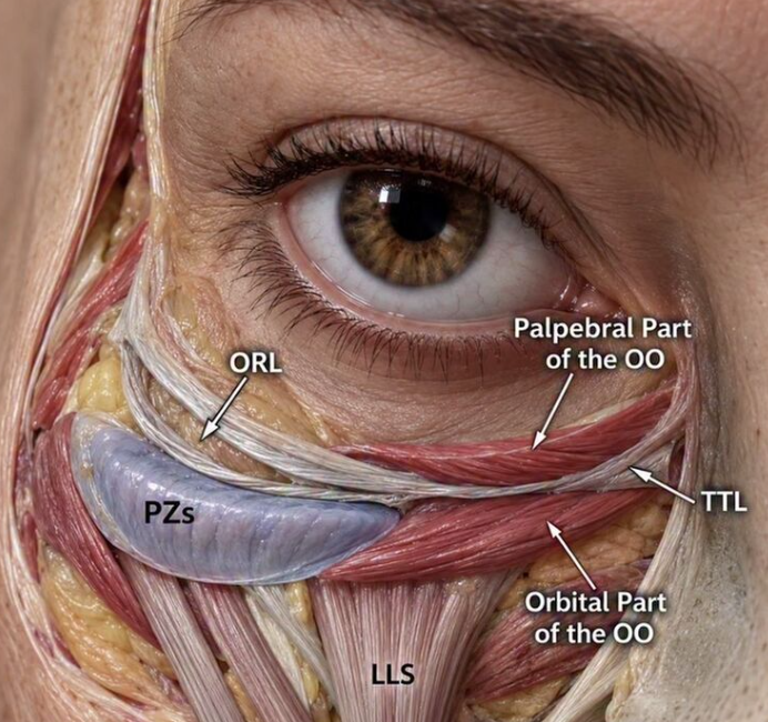
The tear trough is the depression extending downward and outward from the inner corner of the eye. One of its key anatomical foundations is the tear trough ligament, a fibrous attachment connecting the skin to the underlying maxillary bone.
This ligament lies between the palpebral and orbital portions of the orbicularis oculi muscle, the muscle surrounding the eye. Laterally, it continues as the orbicularis retaining ligament, contributing to the attachment between the lower eyelid and cheek.
These attachments help define the contour beneath the eye. As adjacent tissues change in volume or position, the tethered area can become more noticeable. A depression beside a bulge creates a shadow, making the under-eye region appear darker even when skin pigmentation is not the main problem.
Orbital fat and the orbital septum: understanding the bulge
The eye is cushioned by orbital fat. In the lower eyelid, this fat is arranged into medial, central, and lateral compartments. The orbital septum is a thin fibrous layer forming part of the boundary between the orbital contents and the more superficial eyelid tissues.
When orbital fat becomes prominent anteriorly, it can produce visible under-eye bags. A hollow along the infraorbital rim, the lower bony edge of the eye socket, accentuates the contrast between the eyelid and cheek. This transition is called the lid–cheek junction.
The appearance therefore depends on several interacting structures: the fat, retaining ligaments, skin, cheek tissues, and underlying bone.
How these structures combine to create dark circles
Together, prominent orbital fat, the hollow defined by the tear trough ligament, and changes in the surrounding skin and cheek tissues can create an uneven transition between the lower eyelid and cheek.
The bulge catches the light, while the depression beneath it falls into shadow. This combination contributes to what we commonly call under-eye dark circles, sometimes further accentuated by pigmentation or visible blood vessels beneath thin skin. Understanding these overlapping factors helps explain why effective treatment requires assessing the entire lid–cheek junction.
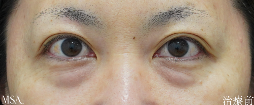
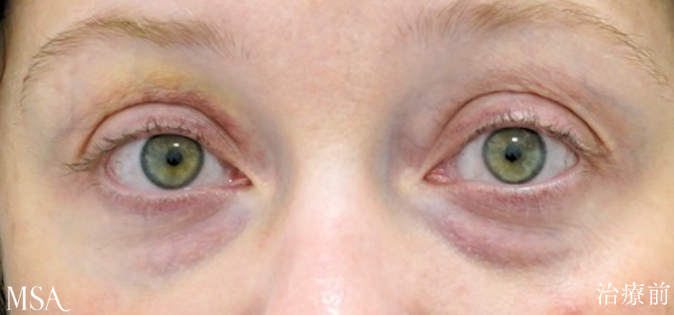
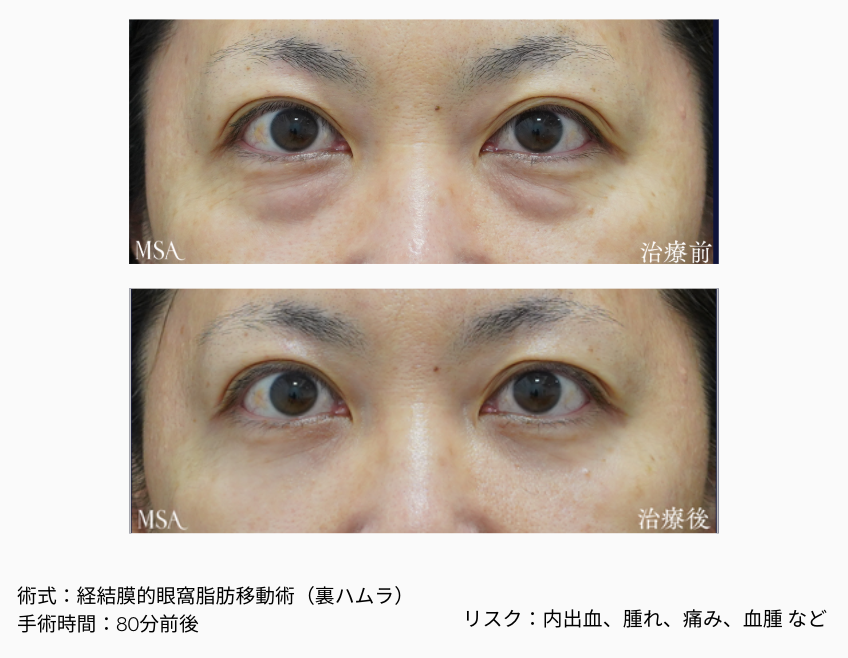
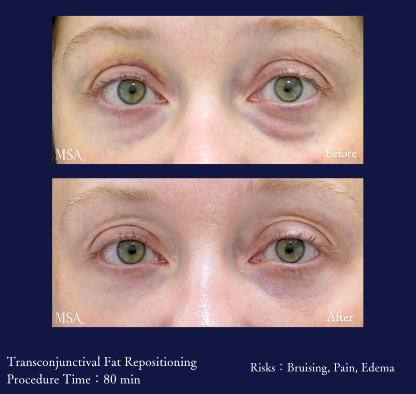
Here are examples of patients with a strong tear trough ligament and bulging fat. You can see how the volume of fat is not the only factor contributing to dark circles ( patient on right has less fat, but circles are prominent ).
How transconjunctival fat repositioning addresses the anatomy
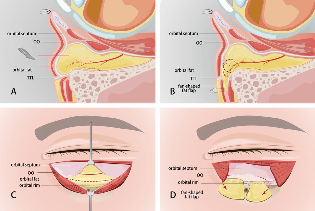
Transconjunctival fat repositioning uses an incision inside the lower eyelid to access the orbital fat.
Where indicated, tethering attachments around the tear trough and orbital rim are released to allow the tissues to redistribute. The fat is then moved over the orbital rim into the hollow beneath it while retaining its attachment to the original tissue. This helps soften the transition between the lower eyelid and cheek.
Another important landmark is the arcus marginalis, the region where the orbital septum joins the periosteum at the orbital rim. Releasing this attachment can permit fat to be redraped across the rim.
The aim is to use existing volume to reduce the contrast between the bulge and the depression. Fat removal alone remains appropriate for some patients, but it may leave an existing tear trough insufficiently corrected.
Septal reset and internal fixation
A septal reset involves advancing and securing the orbital septum over the orbital rim, allowing the septum and associated fat to contribute to a smoother eyelid–cheek contour. It is a distinct maneuver from repositioning individual fat compartments alone.
Internal fixation refers to securing the repositioned tissues with sutures placed internally. The specific release, repositioning, and fixation methods depend on the patient’s anatomy and the surgical plan.
The photographs:
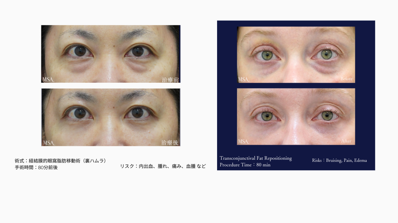
The accompanying photographs show patient before treatment and 1month after transconjunctival fat repositioning.
The postoperative image shows less prominent under-eye bulging and a smoother transition toward the cheeks. Some coloration remains visible, illustrating the distinction between structural shadowing and skin color.
Three weeks is an early postoperative stage. Residual swelling may still influence the contour, so this should be regarded as an early result rather than the final outcome.
Choosing treatment based on anatomy
A careful assessment considers the tear trough, orbital fat, skin quality, eyelid support, and cheek contour together. Fat repositioning does not directly treat every cause of dark circles, and additional treatment may be needed for pigmentation or excess skin.
At MSA Surgical Arts, our goal is to create a smoother, natural-looking transition beneath the eyes while preserving the patient’s individual features.
As with any surgery, potential risks include bruising, swelling, discomfort, dry eyes, asymmetry, contour irregularities, bleeding, infection, and changes in eyelid position. Serious complications affecting vision are rare. Recovery and results vary between patients.
References
Hamra ST. The role of the septal reset in creating a youthful eyelid-cheek complex in facial rejuvenation. Plastic and Reconstructive Surgery. 2004;113(7):2124–2141; discussion 2142–2144. doi: 10.1097/01.PRS.0000122410.19952.E7.
Wong CH, Hsieh MKH, Mendelson B. The tear trough ligament: anatomical basis for the tear trough deformity. Plastic and Reconstructive Surgery. 2012;129(6):1392–1402. doi: 10.1097/PRS.0b013e31824ecd77.
Goldberg RA. Transconjunctival orbital fat repositioning: transposition of orbital fat pedicles into a subperiosteal pocket. Plastic and Reconstructive Surgery. 2000;105(2):743–748; discussion 749–751. doi: 10.1097/00006534-200002000-00044.
reservation診察予約
お気軽にお問い合わせください。
診療時間:9:00~18:00
休診日:不定休
 LINE予約
accessアクセス
MSA美容外科
〒900-0006
沖縄県那覇市おもろまち4-9-10ブギービル5階
- 電話番号:
- 098-867-3311
- 診療科目:
- 美容皮膚科・美容外科・皮膚科・形成外科
- アクセス:
- 那覇メインプレイスから徒歩5分
- 休診日:
- 不定休
- 駐車場:
- 駐車場について
 WEB予約
 電話予約